It is a painless, non-invasive examination that uses high-frequency sound waves (not ionizing X-rays) to image the tissues of the human body, and through which the doctor can make a diagnosis and suggest further treatment. Ultrasound waves travel through the body and reflect off individual organs or transitions between tissues with different acoustic impedances. The disadvantage is the inability of ultrasound to pass through bone and air, making it impossible to examine, for example, the lungs or loops of intestine. During the examination it is necessary to exert sufficient pressure on the measuring probe for good contact with the patient's body, which can be uncomfortable, therefore the surface of the probe is also covered with a gel (ECG cream) to avoid air gaps between the probe and the body of the examined. Because the tissues are imaged in real time during the ultrasound examination, it is possible to capture the structure and movement of the individual organs, as well as the blood flow in the blood vessels.
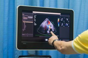
The Doppler effect, described in 1842, is used to measure blood flow. In a Doppler measurement ("colour Doppler"), the relative movement of the blood relative to the probe emitting the ultrasound is displayed by different coloured fields within the sections through the blood vessels or heart. It is therefore possible to detect any narrowing or blockage of blood vessels or, for example, an atrial defect in the heart.
Harmful effects of the examination?
No adverse side effects have been demonstrated during the time ultrasound has been used in medicine, but it should only be used for clear indications, on technically suitable equipment, and should always be performed by a qualified physician.
Indications for an ultrasound examination
The examination is performed to assess the morphological findings in the area of interest. In particular, it is an assessment of the organs in the abdominal cavity, in the soft tissues of the neck (thyroid gland) and elsewhere on the body. Using the Doppler modality, it is possible to assess the patency of the blood vessels and the filling of the cardiac compartments. As mentioned above, bone tissue and air-filled organs (lung parenchyma) cannot be examined.
Reduced yield of ultrasound examinations
- An uncooperative patient.
- Obesity.
- Flatulence.
- Adverse anatomical conditions.
- Calcification.
Appointment for examination
The examination is ordered by the attending physician for both inpatients and outpatients. As with other examinations, priority is given to patients in serious clinical condition.
Patient preparation
The examination is performed lying down (on the back or side).
Ultrasound of the abdominal organs - the patient presents for this examination on an empty stomach (except in acute cases).
Ultrasound of the bladder with residue - here it is necessary for the patient to have a full bladder, it is advisable to drink 0.5 - 1 litre of fluid about 1 hour before the examination.
UZ of other organs - without preparation.
You can make an appointment / inquire about an examination by calling 567 157 602.
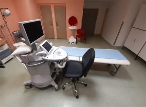
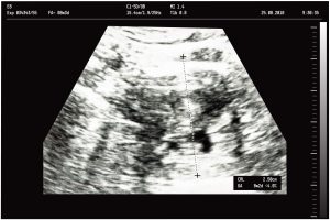
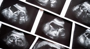
Instrumentation and sample images:
Leave a Reply
You must be logged in to post a comment.